Anterior Pituitary
The anterior adenophypophysis is divided into the pars tuberalis, pars intermedia and pars distalis.
The pars intermedia (the intermediate lobe) lies between the pars distalis and the pars tuberalis, and is rudimentary in the humans
Endocrine cells of the anterior pituitary are controlled by regulatory hormones released by parvocellular neurosecretory cells in the hypothalamic capillaries leading to infundibular blood vessels, which in turn lead to a second capillary bed in the anterior pituitary.
This vascular relationship constitutes the hypothalamo-hypophyseal portal system. Diffusing out of the second capillary bed, the hypothalamic releasing hormones then bind to anterior pituitary endocrine cells, upregulating or downregulating their release of hormones.[4]
The anterior pituitary contains several different types of cells[5] that synthesize and secrete hormones. Usually there is one type of cell for each major hormone formed in anterior pituitary. With special stains attached to high-affinity antibodies that bind with distinctive hormone, at least 5 types of cells can be differentiated.
| Type of cell | Hormone secreted |
|---|---|
| Somatotropes | human growth hormone (hGH) |
| Corticotropes | adrenocorticotropin (ACTH) |
| Thyrotropes | thyroid stimulating hormone (TSH) |
| Gonadotropes | gonadotropic hormone i.e., both luteinizing hormone (LH) and follicle stimulating hormone (FSH) |
| Lactotropes | prolactin (PRL) |
Each secretory cell in the adenohypophysis synthesizes and stores one of the following hormones: follicle-stimulating hormone (FSH), thyrotropin (thyroid-stimulating hormone [TSH]), luteinizing hormone (LH), adrenocorticotropic hormone (ACTH), growth hormone (GH), or prolactin. These hormones control the secretory activities of many other glands. Their release is regulated by specific releasing or inhibiting hormones produced by the hypothalamus and delivered to the adenohypophysis by the blood in the hypophyseal portal system (III.D).
-
Chromophobes. These cells stain poorly and appear clear or white in tissue sections. Together, the three chromophobe subpopulations make up approximately 50% of the pars anterior’s epithelial cells. They include (1) undifferentiated nonsecretory cells, which may be stem cells; (2) partly degranulated chromophils, which contain sparse granules; and (3) follicular cells, the predominant chromophobe type, which form a stromal network supporting the chromophils; these stellate cells may be phagocytic.
-
Chromophils. These hormone-secreting cells stain intensely, owing to the abundant cytoplasmic secretory granules in which hormones are stored. A specific cell type exists for each hormone. Usually larger than chromophobes, chromophils comprise two classes:
-
Acidophils secrete simple proteins and stain intensely with eosin but respond negatively to the PAS reaction. More abundant in the gland periphery, they are usually smaller than basophils and have larger and more numerous granules. The acidophils include two major hormone-secreting cell types: somatotrophs produce GH, (somatotropin) and mammotrophs produce prolactin. (A mnemonic for hormones secreted by acidophils is GPA: growth hormone, prolactin, acidophils.)
-
Basophils secrete glycoproteins, stain with hematoxylin and other basic dyes, and respond positively to the PAS reaction. More abundant in the core of the gland, they are usually larger than acidophils, with fewer and smaller granules. The three major hormone-producing basophils produce four major hormones. (A mnemonic for hormones produced by basophils is B-FLAT: basophils, FSH, LH, ACTH, TSH.) Each of the two gonadotrophs produces a different gonadotropin. One produces FSH; the other produces LH. Corticotrophs produce adrenocorticotropin (ACTH). Thyrotrophs produce thyrotropin (TSH).
-
This funnel-shaped superior extension of the pars distalis surrounds the infundibular stem (Fig. 20–2). It resembles the pars distalis but contains mostly gonadotrophs. The pars tuberalis contains many capillaries of the primary capillary plexus (III.D.1).
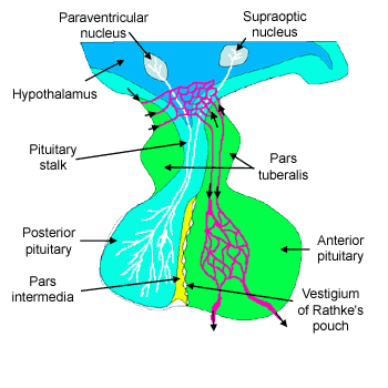
FIGURE 20–2.
Diagram of the subdivisions, blood supply, and innervation of the pituitary gland and hypothalamus. The adenohypophysis (gray, left) lies anterior to the neurohypophysis (right). For simplicity, only a few of the many nuclei of the hypothalamus (top) are shown. Major subdivisions are shown in boldface.

This is a band or wedge of adenohypophysis between the pars distalis and pars nervosa; it is rudimentary in humans. It contains Rathke’s cysts—small, irregular, colloid-containing cavities lined with cuboidal epithelium that are the remnants of Rathke’s pouch. It also contains scattered clumps and cords of basophilic cells, or melanotrophs, which secrete melanocyte-stimulating hormone (β-MSH).
The pituitary gland is at the base of the brain, about 1cm in diameter, lying beneath the third ventricle in a bony cavity (sella turcica) in the base of the skull. (See Gross Anatomy: Pituitary Gland

III. Adenohypophysis
Each secretory cell in the adenohypophysis synthesizes and stores one of the following hormones: follicle-stimulating hormone (FSH), thyrotropin (thyroid-stimulating hormone [TSH]), luteinizing hormone (LH), adrenocorticotropic hormone (ACTH), growth hormone (GH), or prolactin. These hormones control the secretory activities of many other glands. Their release is regulated by specific releasing or inhibiting hormones produced by the hypothalamus and delivered to the adenohypophysis by the blood in the hypophyseal portal system (III.D).
-
Chromophobes. These cells stain poorly and appear clear or white in tissue sections. Together, the three chromophobe subpopulations make up approximately 50% of the pars anterior’s epithelial cells. They include (1) undifferentiated nonsecretory cells, which may be stem cells; (2) partly degranulated chromophils, which contain sparse granules; and (3) follicular cells, the predominant chromophobe type, which form a stromal network supporting the chromophils; these stellate cells may be phagocytic.
-
Chromophils. These hormone-secreting cells stain intensely, owing to the abundant cytoplasmic secretory granules in which hormones are stored. A specific cell type exists for each hormone. Usually larger than chromophobes, chromophils comprise two classes:
-
Acidophils secrete simple proteins and stain intensely with eosin but respond negatively to the PAS reaction. More abundant in the gland periphery, they are usually smaller than basophils and have larger and more numerous granules. The acidophils include two major hormone-secreting cell types: somatotrophs produce GH, (somatotropin) and mammotrophs produce prolactin. (A mnemonic for hormones secreted by acidophils is GPA: growth hormone, prolactin, acidophils.)
-
Basophils secrete glycoproteins, stain with hematoxylin and other basic dyes, and respond positively to the PAS reaction. More abundant in the core of the gland, they are usually larger than acidophils, with fewer and smaller granules. The three major hormone-producing basophils produce four major hormones. (A mnemonic for hormones produced by basophils is B-FLAT: basophils, FSH, LH, ACTH, TSH.) Each of the two gonadotrophs produces a different gonadotropin. One produces FSH; the other produces LH. Corticotrophs produce adrenocorticotropin (ACTH). Thyrotrophs produce thyrotropin (TSH).
-
This funnel-shaped superior extension of the pars distalis surrounds the infundibular stem (Fig. 20–2). It resembles the pars distalis but contains mostly gonadotrophs. The pars tuberalis contains many capillaries of the primary capillary plexus (III.D.1).
FIGURE 20–2.
Diagram of the subdivisions, blood supply, and innervation of the pituitary gland and hypothalamus. The adenohypophysis (gray, left) lies anterior to the neurohypophysis (right). For simplicity, only a few of the many nuclei of the hypothalamus (top) are shown. Major subdivisions are shown in boldface.
This is a band or wedge of adenohypophysis between the pars distalis and pars nervosa; it is rudimentary in humans. It contains Rathke’s cysts—small, irregular, colloid-containing cavities lined with cuboidal epithelium that are the remnants of Rathke’s pouch. It also contains scattered clumps and cords of basophilic cells, or melanotrophs, which secrete melanocyte-stimulating hormone (β-MSH).